![[Figure 1A]](http://images.squarespace-cdn.com/content/v1/58e61af4a5790ab156e17bc1/04fe6ec5-55ff-401c-bf64-4d2b756ff838/Logo+transparente+background.png)
GALLERY | finalists OF THE [FIGURE 1.A.] 2018 EXHIBITION
The second edition of the [Figure 1.A.] exhibition invited researchers from the University of Lausanne's Faculty of Biology and Medicine (FBM) to send their images. The jury chose 58 photographs to be shown in the Gallery La Sonnette in downtown Lausanne.
Below is a list of the jury and audience favourites. Scroll down to read the description and authors’ names.
JURY AWARDS

Arabidopsis Diatom
Pauline Anne | Department of Plant Molecular Biology, UNIL, Switzerland
Artistic arrangement of Arabidopsis thaliana roots expressing a fluorescent marker.
FR: Arrangement artistique de racines d'Arabidopsis thaliana exprimant un marqueur fluorescent.
![09_48x60cm_[objectofscience_2]_[nusbaumer_david]_[comet+appocalypse].jpeg](https://images.squarespace-cdn.com/content/v1/58e61af4a5790ab156e17bc1/1621247207884-B6RKBAX28XDNYWU471EZ/09_48x60cm_%5Bobjectofscience_2%5D_%5Bnusbaumer_david%5D_%5Bcomet%2Bappocalypse%5D.jpeg)
Comet Apocalypse
David Nusbaumer | Department of Ecology and Evolution, UNIL, Switzerland
This is a microscopy image of nuclei of the spermatozoa of the Arctic char (Salvelinus alpinus) during a so-called comet assay. The DNA is stained and if it is intact it will be seen as a ball. However, when there is damage to the DNA, little fragments appear and form the comet tail. This image was taken as a part of a project on how female char choose their partners, especially under intense male competition and in the presence of parasitic spawning.
FR: Voici une image de microscopie des noyaux des spermatozoïdes de l'omble chevalier (Salvelinus alpinus) au cours d'un test dit des comètes. L'ADN est coloré et s'il est intact, on le voit comme une boule. Cependant, lorsque l'ADN est endommagé, de petits fragments apparaissent et forment la queue de la comète. Cette image a été prise dans le cadre d'un projet sur la manière dont les ombles femelles choisissent leurs partenaires, notamment en cas de concurrence intense entre les mâles et en présence d'une ponte parasitaire.
Find more of David’s photography here www.kiwiphotography.ch and on Instagram @kiwiphotography.ch

Abstract Development
Vittoria Mariano | Department of Fundamental Neuroscience, UNIL, Switzerland
The making of a wing and a leg: this is a microscopy picture representing 2 sac-like structures that are found inside the larva of a fly, undergoing metamorphosis. These sacs are called the Imaginal Discs: the small round one will transform into a leg, while the bigger one will transform into the wing.
FR: La fabrication d'une aile et d'une patte : il s'agit d'une image de microscopie représentant 2 structures en forme de sac que l'on trouve à l'intérieur de la larve d'une mouche, en cours de métamorphose. Ces sacs sont appelés les disques imaginaux : le petit rond se transformera en patte, tandis que le plus grand se transformera en aile.

Morille
Nathalie Busso and Véronique Chobaz | Service de Rhumatologie, CHUV, Switzerland
This is a histological staining from a section from the knee joint of a mouse, in which you can see the cartilage growth plate.
FR: Il s'agit d'une coloration histologique d'une section de l'articulation du genou d'une souris, dans laquelle vous pouvez voir la plaque de croissance du cartilage.

Neurons in a box
Nicolas Toni | Centre de Neurosciences Psychiatriques, CHUV, Switzerland
In the brain, adult neural stem cells continuously generate neurons. This process is named adult neurogenesis and is crucial for memory and resistance to depression. By growing these cells in a Petri dish, one can easily manipulate them and hopefully, understand how they can be pushed to generate more neurons for potential therapeutic applications. This is a microscopy picture of neurons (yellow) and adult neural stem cells (purple), in a Petri dish. Image size: 200x160 micrometres.
FR: Dans le cerveau, les cellules souches neurales adultes génèrent continuellement des neurones. Ce processus, appelé neurogenèse adulte, est crucial pour la mémoire et la résistance à la dépression. En cultivant ces cellules dans une boîte de Pétri, on peut facilement les manipuler et, espérons-le, comprendre comment les pousser à générer davantage de neurones pour des applications thérapeutiques potentielles. Image microscopique de neurones (en jaune) et de cellules souches neurales adultes (en violet), dans une boîte de Pétri. Taille de l'image : 200x160 micromètres
AUDIENCE FAVOURITES

Lonely in the dark
David Nusbaumer | Department of Ecology and Evolution, UNIL, Switzerland
The European grayling from Lake Thun are under strong decline and are endangered. In this picture, the author wanted to illustrate the drastic population decline by photographing this lonely embryo on the gravel bed.
FR: Les ombres européens du lac de Thoune connaissent un fort déclin et sont en voie de disparition. Dans cette photo, l'auteur a voulu illustrer le déclin drastique de la population en photographiant cet embryon solitaire sur le lit de gravier.
Find more of David’s photography here www.kiwiphotography.ch and on Instagram @kiwiphotography.ch

millimeter
Joel Wellbourne-Wood | Department of Fundamental Neuroscience, UNIL, Switzerland
Since most single cells are invisible to the naked eye, conceptualizing their size in your mind can be difficult. In order to circumvent this, the author has pictured a single square millimetre of space, containing bovine pulmonary artery endothelial cells. Since 1 mm can be resolved with the naked eye this photo provides a reference upon which the viewer can judge their individual size. The cells’ cytoskeleton is stained green. Their mitochondria, which provide energy to the cell, are stained in red. The nucleus, which contains the cells’ DNA, is stained in blue.
FR: Comme la plupart des cellules uniques sont invisibles à l'œil nu, il peut être difficile de conceptualiser leur taille dans votre esprit. Afin de contourner ce problème, l'auteur a photographié un seul millimètre carré d'espace, contenant des cellules endothéliales d'artères pulmonaires bovines. Comme un millimètre peut être résolu à l'œil nu, cette photo fournit une référence à partir de laquelle le spectateur peut juger de leur taille individuelle. Le cytosquelette des cellules est coloré en vert. Leurs mitochondries, qui fournissent l'énergie à la cellule, sont colorées en rouge. Le noyau, qui contient l'ADN des cellules, est coloré en bleu.
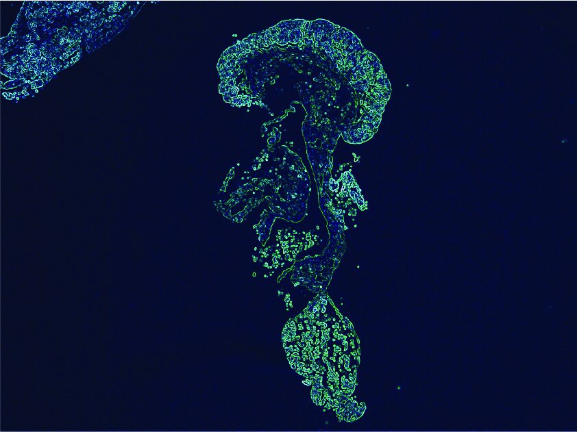
jellyfish
Lucie Vivancos | Département Femme Mère Enfant, CHUV, Switzerland
A microscopy image of a piece of a neuroblastoma tumour.
FR: Image microscopique d'un morceau d'une tumeur de neuroblastome.
Hands at work 2018
Cassandre Kinnaer | Department of Fundamental Microbiology, UNIL, Switzerland
BEFORE WINTER
Biologists working with honey bees (Apis mellifera carnica) have to make sure the hive is healthy including that the queen is normally laying eggs. They do this once a week and it is especially important to check the well-being of the bees before winter when they will start hibernating to survive the cold temperatures.
FR: Les biologistes qui travaillent avec des abeilles domestiques (Apis mellifera carnica) doivent s'assurer que la ruche est en bonne santé et que la reine pond normalement. Ils le font une fois par semaine et il est particulièrement important de vérifier le bien-être des abeilles avant l'hiver, lorsqu'elles commenceront à hiberner pour survivre aux températures froides.
ENTROPY
Bio-mathematicians use equations to model diverse biological phenomenon. On the picture we can see part of an entropy computation that is here applied to model DNA strand behaviour, deepening our understanding of this complex molecule that supports life as we know it.
FR: Les bio-mathématiciens utilisent des équations pour modéliser divers phénomènes biologiques. Sur l'image, nous pouvons voir une partie d'un calcul d'entropie qui est ici appliqué pour modéliser le comportement des brins d'ADN, approfondissant ainsi notre compréhension de cette molécule complexe qui soutient la vie telle que nous la connaissons.
THE CREATION 2.0
Depicted here is a caterpillar from the species Pieris brassicae and it is carried on the hand by a scientist. This species can cause important damage to crops and it is, therefore, important to understand the relationship between plant and insect and how plants defend themselves against aggression by herbivores.
FR: La photo représente une chenille de l'espèce Pieris brassicae et elle est portée à la main par un scientifique. Cette espèce peut causer des dégâts importants sur les cultures et il est donc important de comprendre la relation entre la plante et l'insecte et comment les plantes se défendent contre les agressions des herbivores.
FOUR BY FOUR
The white dots are growing colonies of fission yeast Schizosaccharomyces pombe. After the sexual cycle, S. pombe will produce four spores or offspring. Each colony originates from a single spore which was placed on the petri dish with the help of a dissector, a machine with a very small needle that can pick a single cell. Biologists use this tool to perform very thorough genetic analyses.
FR: Les points blancs sont des colonies en croissance de la levure de fission Schizosaccharomyces pombe. Après le cycle sexuel, S. pombe produira quatre spores ou progénitures. Chaque colonie provient d'une seule spore qui a été placée sur la boîte de Pétri à l'aide d'un dissecteur, une machine dotée d'une très petite aiguille qui peut prélever une seule cellule. Les biologistes utilisent cet outil pour effectuer des analyses génétiques très poussées.

This work is licensed under a Creative Commons Attribution-NonCommercial-NoDerivatives 4.0 International License.