![[Figure 1A]](http://images.squarespace-cdn.com/content/v1/58e61af4a5790ab156e17bc1/04fe6ec5-55ff-401c-bf64-4d2b756ff838/Logo+transparente+background.png)
GALLERY | finalists OF THE [FIGURE 1.A.] 2017 EXHIBITION
The first edition of the [Figure 1.A.] exhibition invited researchers from the University of Lausanne's Faculty of Biology and Medicine (FBM) to send their images. The jury chose 58 photographs to be shown in the Gallery La Sonnette in downtown Lausanne.
Below is a list of the jury and audience favourites. Click on images to read the description and authors’ names.
JURY AWARDS

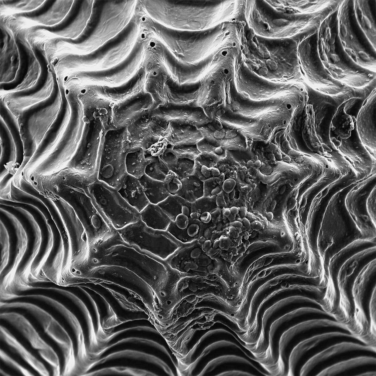
PATTERNING
Raphaël Groux, Antonio Mucciolo | Department of Plant Molecular Biology and Electron microscopy facility, UNIL, Switzerland
560x magnification of a Pieris brassicae egg. Insect eggs display a wonderful degree of symmetry and the presence of aeropyles ("holes" through which the egg breathes). Hiding within this egg is one of the most feared enemies of plants: a caterpillar.
FR: Agrandissement (560x) d’un oeuf de la piéride du chou. Les oeufs des insectes montrent un grand degré de symétrie et la présence d’aéropyles, des trous qui permettent à l’oeuf de respirer. Dans ces oeufs se cache l’un des plus grands ennemis des plantes : une chenille.

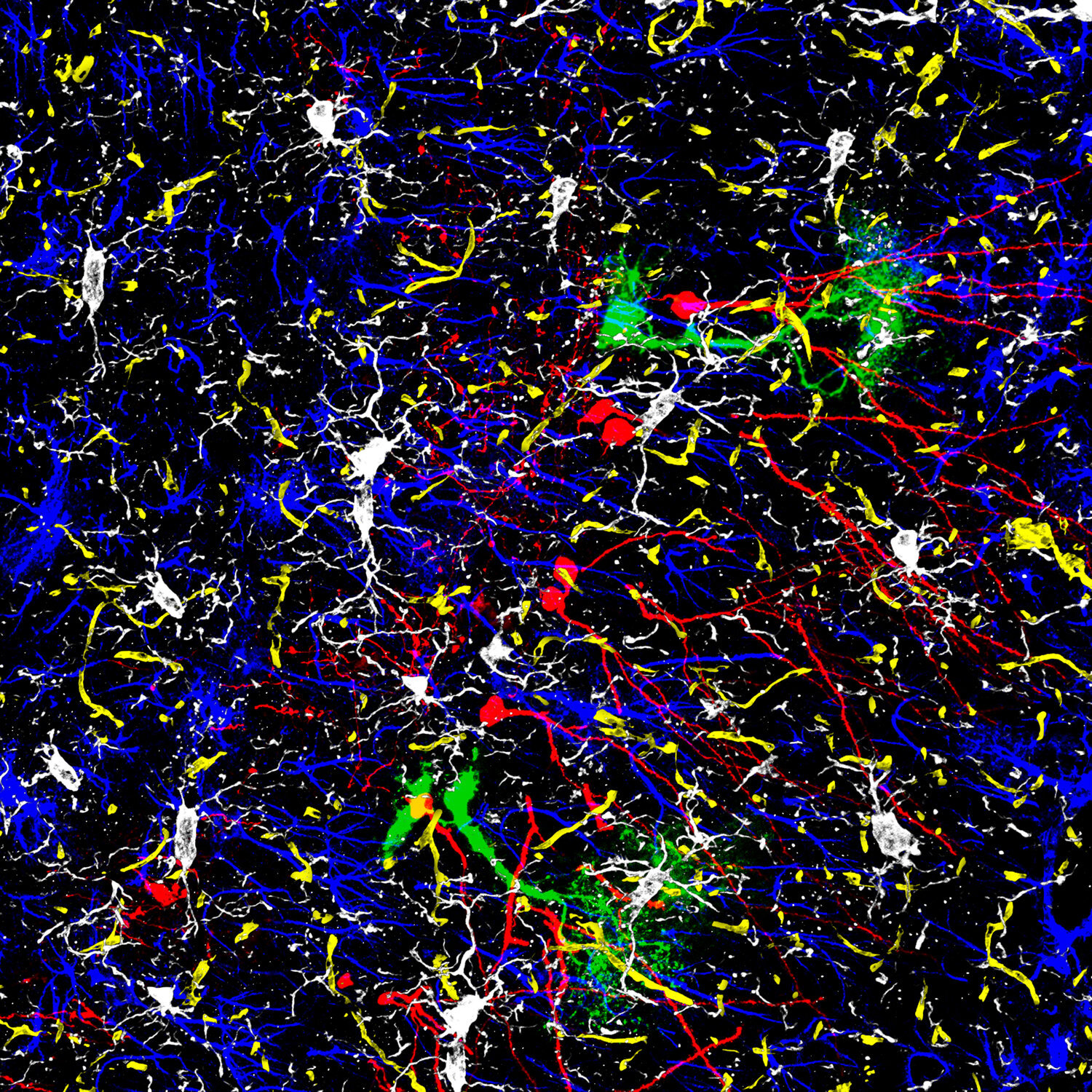
The Brain Crowd
Elias Gebara, Nicolas Toni | Centre of Psychiatric Neuroscience, CHUV, Switzerland
ALSO AUDIENCE FAVOURITE
The brain is generally considered a network of neurons. This image shows the diversity of cells that also constitute it: Astrocytes (blue), microglia (white), stem cells (green), blood vessels (yellow) and some immature neurons (red). The complexity of this picture nicely represents the complexity of the interactions occurring in real-time in the brain.
FR: Le cerveau est généralement considéré comme le réseau de neurones. Cette image montre la diversité des cellules qui le composent : Astrocytes (bleu), la microglie (blanc), cellules souches (verts), les vaisseaux sanguins (jaunes) et certains neurones immatures (rouge). La complexité de cette photo représente bien la complexité des interactions qui se produisent en temps réel dans le cerveau.

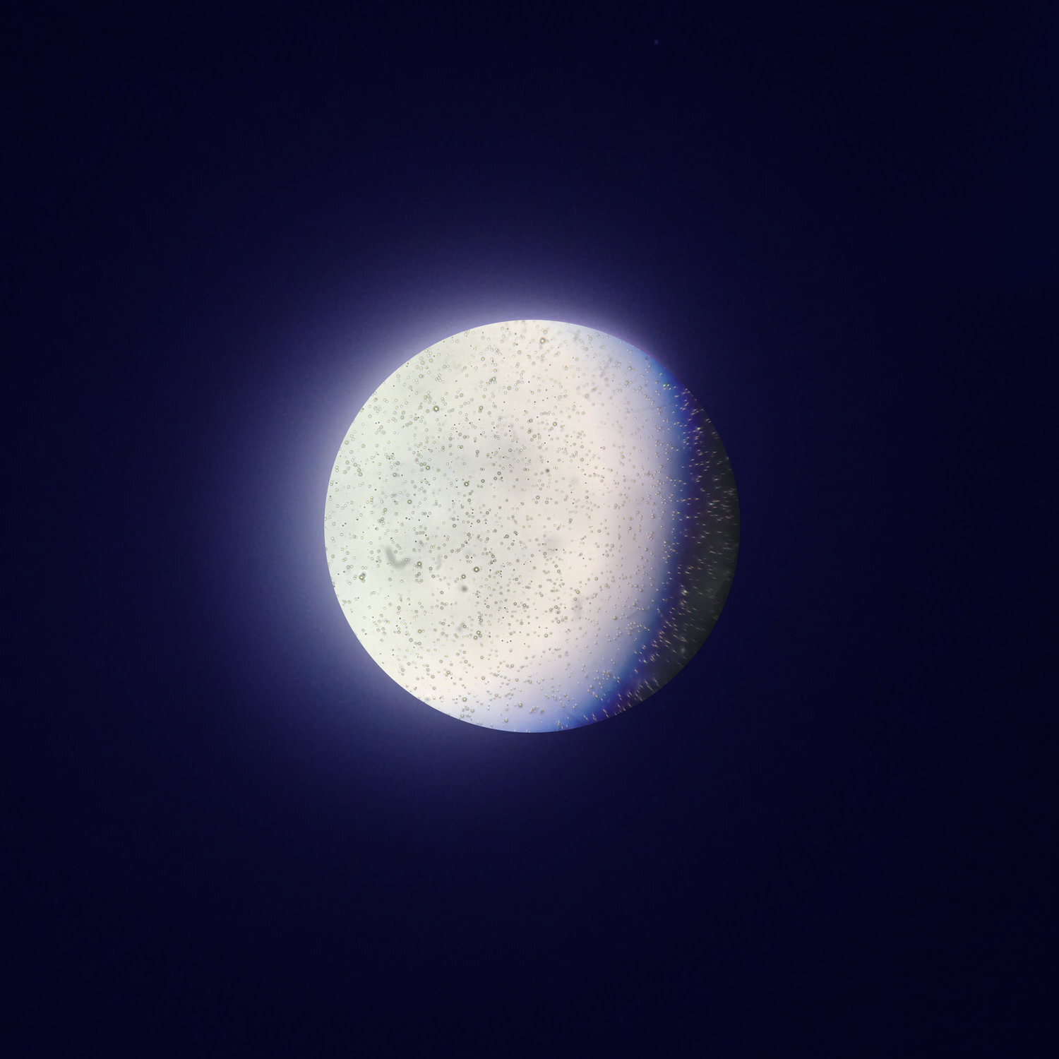
Crescent Mom at Work
Silvia Fuertes Marraco | Department of Fundamental Oncology, UNIL, Switzerland
Breastmilk is alive and cycles through its lifetime: in addition to all the incomparable and evolving nutrients it contains vast amounts of immune cells that protect the mother and infant dyad. We now know that breastmilk also contains stem cells that may incorporate in the infant’s organs, providing yet unrevealed functions. Silvia is an immunologist that came back to the lab from her maternity leave and continued to breastfeed her twins: mom is at work, growing (crescent), physically in the lab as well as through her continued production of milk, her career and motherhood. This picture was taken by curiosity, diluting her breastmilk 30 times and staining with trypan blue for eventual dead cell exclusion, using a microscope and an iPhone camera on top of the eyepiece. The round microscope field (moon) is an intact image of what can be seen - the blue shadow that mimics the crescent’s shadow is an optical effect of finding the focus while aligning the camera to the eyepiece. The outside of the round microscope field is digitally edited to have an aura, using the aura of an original photograph of the lunar eclipse in September 2015 and an indigo background.
FR: Le lait maternel est vivant et en constate évolution : en plus des nutriments incomparables, le lait contient une vaste quantité de cellules immunitaires qui protègent la mère et l’enfant. Nous savons aujourd’hui aussi que le lait contient des cellules souches qui pourraient s’incorporer dans les organes de l’enfant allaité, apportant des fonctions encore pas révélés. Silvia est une chercheuse immunologiste qui a repris le travail après son congé maternité et a continué d’allaiter ses jumeaux, des lors: maman est au travail, en croissance, physiquement au laboratoire ainsi que par sa production de lait continue, sa carrière et sa maternité. Cette photo a été prise par curiosité, en diluant son lait 30 fois dans du PBS et avec de la coloration bleu de trypan pour exclure les cellules éventuellement mortes, en utilisant un microscope et une caméra iPhone sur l’oculaire du microscope. Le champ rond (lune) est une image intacte de ce qui est visible au microscope – l’ombre bleu qui imite l’ombre du croissant est un effet optique lorsque qu’on essaye d’aligner la camera sur l’oculaire. L’extérieur du champ rond du microscope est édité digitalement pour ajouter une aura, en utilisant l’aura d’une photo originale de l’éclipse lunaire de septembre 2015 et un fond indigo.

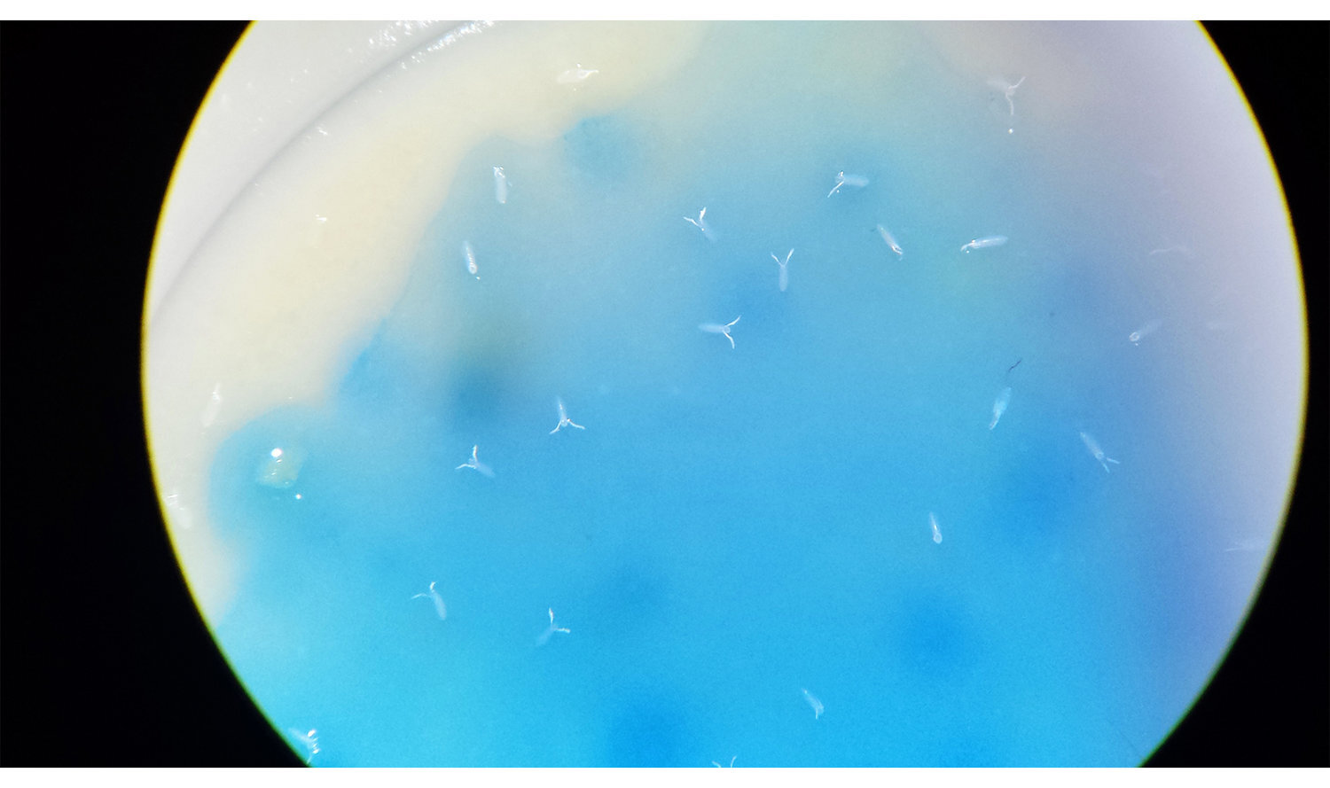
Boats on the Ocean Shore
Kathrin Garschall | Department of Ecology and Evolution, UNIL, Switzerland
This image is from an investigation on whether infecting female fruit flies affects their egg-laying. In this picture, you can see the eggs that one female laid within 24h. The blue dye is a kitchen food colourant that helps to easier spot the white eggs on the fly medium. This image is a result of a mistake the scientist made – she added too little of the colouring dye and the fruit fly eggs suddenly appeared as tiny boats floating in the ocean.
FR: Cette image provient d’une étude qui cherche à déterminer si une infection des femelles drosophiles a une influence sur la ponte de leurs oeufs. Sur cette image, on peut voir les oeufs qu’une femelle a pondus en 24 heures. Le colorant bleu est un colorant alimentaire qui aide à repérer plus facilement les oeufs, blancs, sur le milieu de culture. Cette image résulte d’une erreur commise par la scientifique : elle a utilisé trop peu de colorant, et les oeufs de drosophiles s’apparentent soudainement à de petits bateaux, flottant sur l’océan.

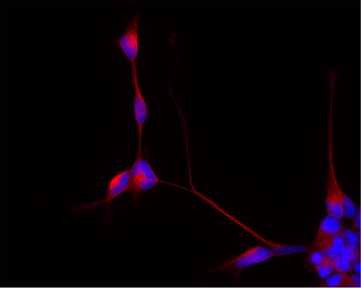
Flowering
Lucie Vivancos | Département femme-mère-enfant, CHUV, Switzerland
Connections between neuronal cells.
FR: Connections entre des cellules neuronales.

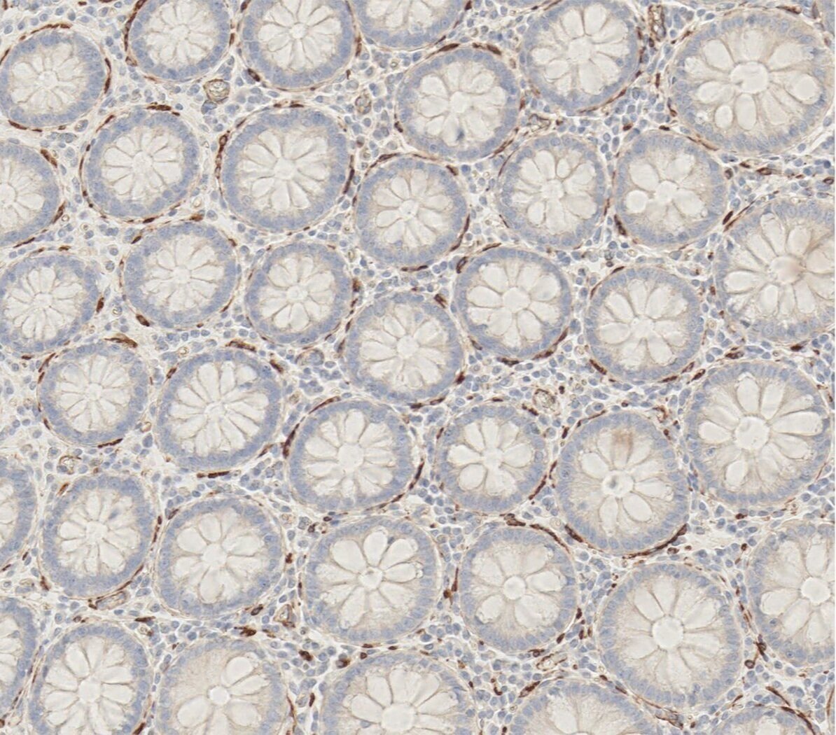
Tentacle Flowers
Christophe Cisarovsky | Department of Fundamental Oncology, CHUV, Switzerland
The surface of the human colon tissue. Each gland stands at an equal distance to its neighbours and is protected and maintained by smooth muscle cells. In imaging it is not that frequent, especially for human and colonic tissue, to obtain a perfect cross-section and appreciate the beauty of cellular organisation. The picture was obtained in a project on colorectal cancer.
FR: La surface du côlon humain. Chaque glande se tient à une distance égale de ses voisines et est protégée et maintenue par des cellules de muscle lisse. Lorsque l’on prend des images, il n’est pas si fréquent, en particulier pour des tissus humains et de côlon, d’obtenir une coupe parfaite et de pouvoir admirer la beauté de l’organisation des cellules. Cette image a été obtenue lors d’un projet étudiant le cancer colorectal.

alien
Alexandros Kanellopoulos, Mariachiara Bucarelli, Vittoria Mariano, Claudia Bagni | Department of Fundamental Neuroscience, UNIL, Switzerland
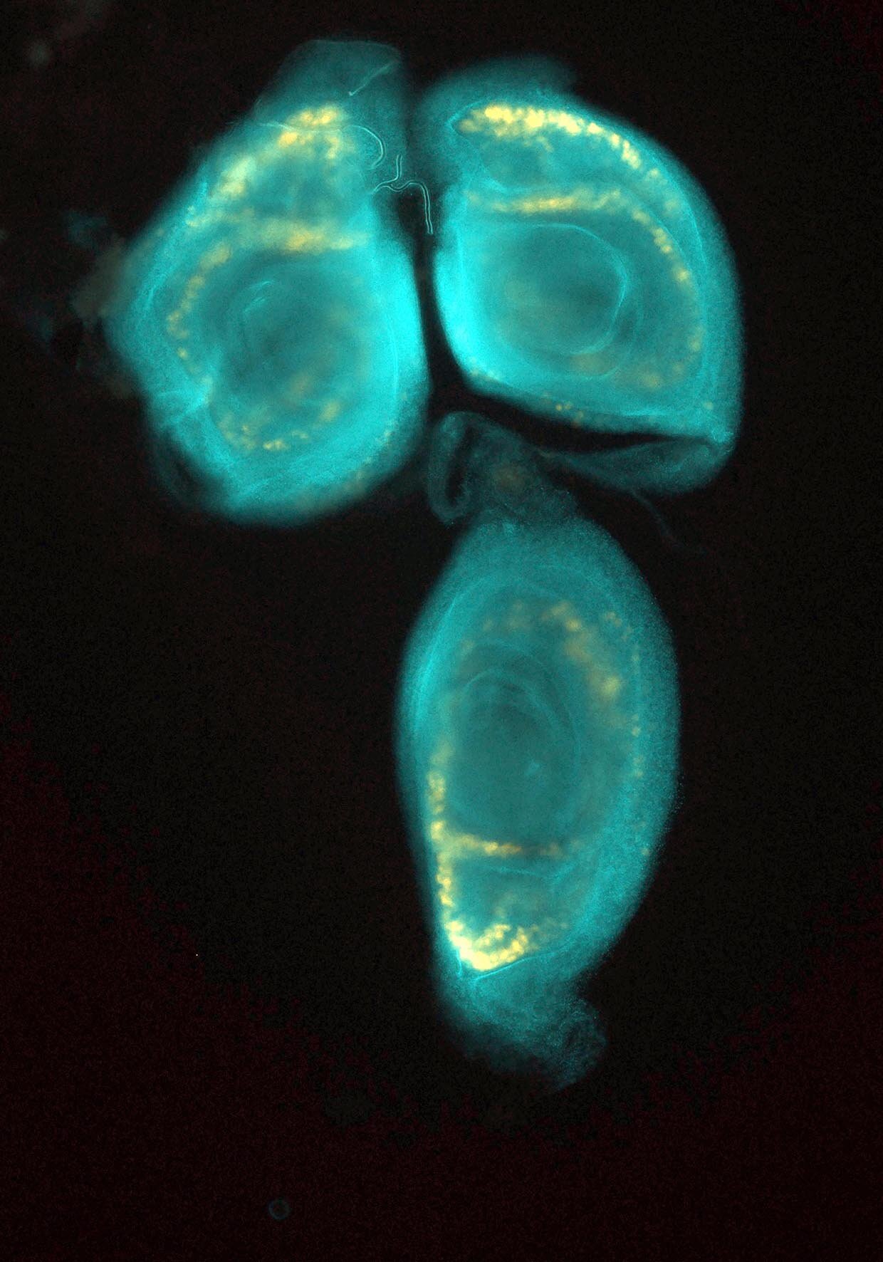
Imaginal discs are the parts of an insect larva that will become a portion of the outside of the adult insect during the pupal transformation. Contained within the body of the larva, they will form the eyes, the wings, the legs, the antennae or other structures in the adult. The role of the imaginal disc in insect development is really important and studying it helps also for investigations of different human diseases, for example, tumour development.
FR: Les disques imaginaux sont des parties de la larve d’insecte qui vont faire partie de l’extérieur de l’insecte après la transformation de la pupe. Contenus dans le corps de la larve, ils formeront les yeux, les ailes, les antennes et d’autres structures de l’insecte adulte. Le rôle des disques imaginaux dans le développement de l’insecte est très important et les étudier peut également aider à la compréhension de différentes maladies humaines, comme par exemple le développement des tumeurs.

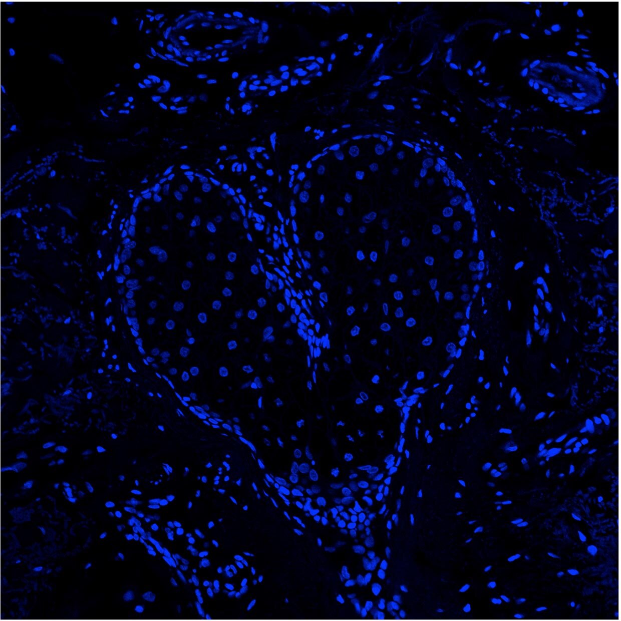
Tough Love
Dania Al Labban | Department of Biochemistry, UNIL, Switzerland
Actinic keratosis is a pre-malignant form of skin cancer. This image shows tissue of a patient who has this form of cancer. While imaging the sample for a gene of interest together with a marker showing the nuclei of cells, the scientist observed this heart. Beautiful patterns can emerge even from dark and scary places.
FR: La kératose actinique est une forme prémaligne de cancer de la peau. Cette image montre le tissu d’un patient qui souffre de cette forme de cancer. En prenant les images d’un échantillon d’un gène d’intérêt avec un marqueur montrant le noyau des cellules, la scientifique a observé ce coeur. De beaux motifs peuvent émerger même d’endroits sombres et effrayants.

Fibers
Berra Erkosar | Department of Ecology and Evolution, UNIL, Switzerland
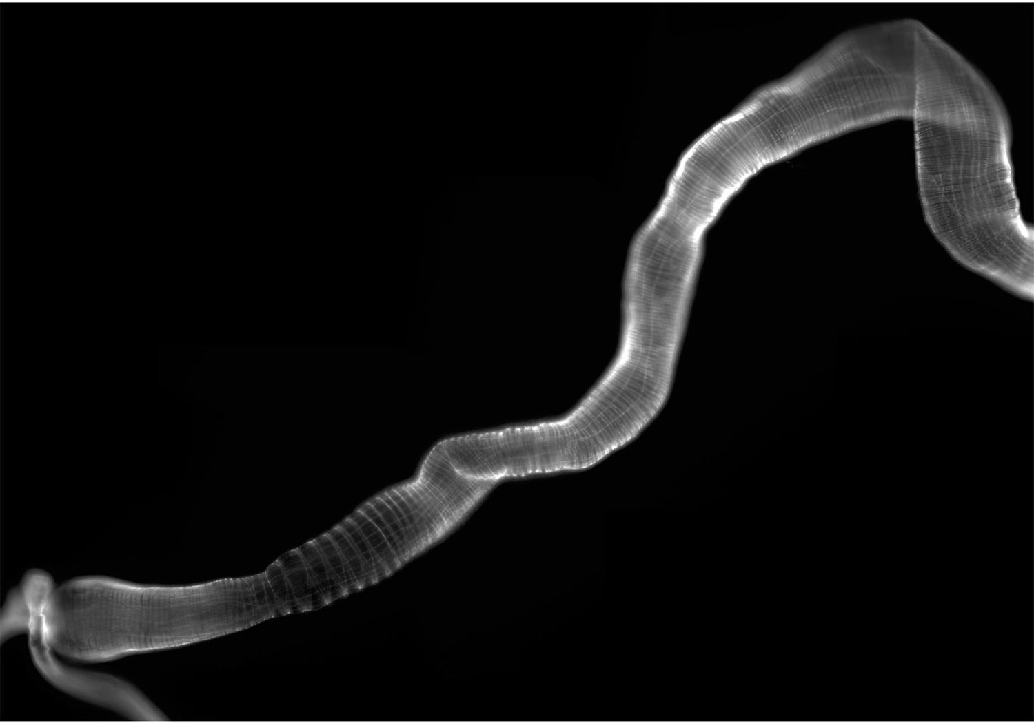
The intestinal muscle structure of the fruit fly Drosophila, where muscle fibers are smoothly and clearly marked.
FR: La structure du muscle intestinal de la Drosophile, où les fibres musculaires sont sans à-coup et clairement marquées.

Gazing Inside
Irina Kolotueva | Electron Microscopy Facility, UNIL, Switzerland
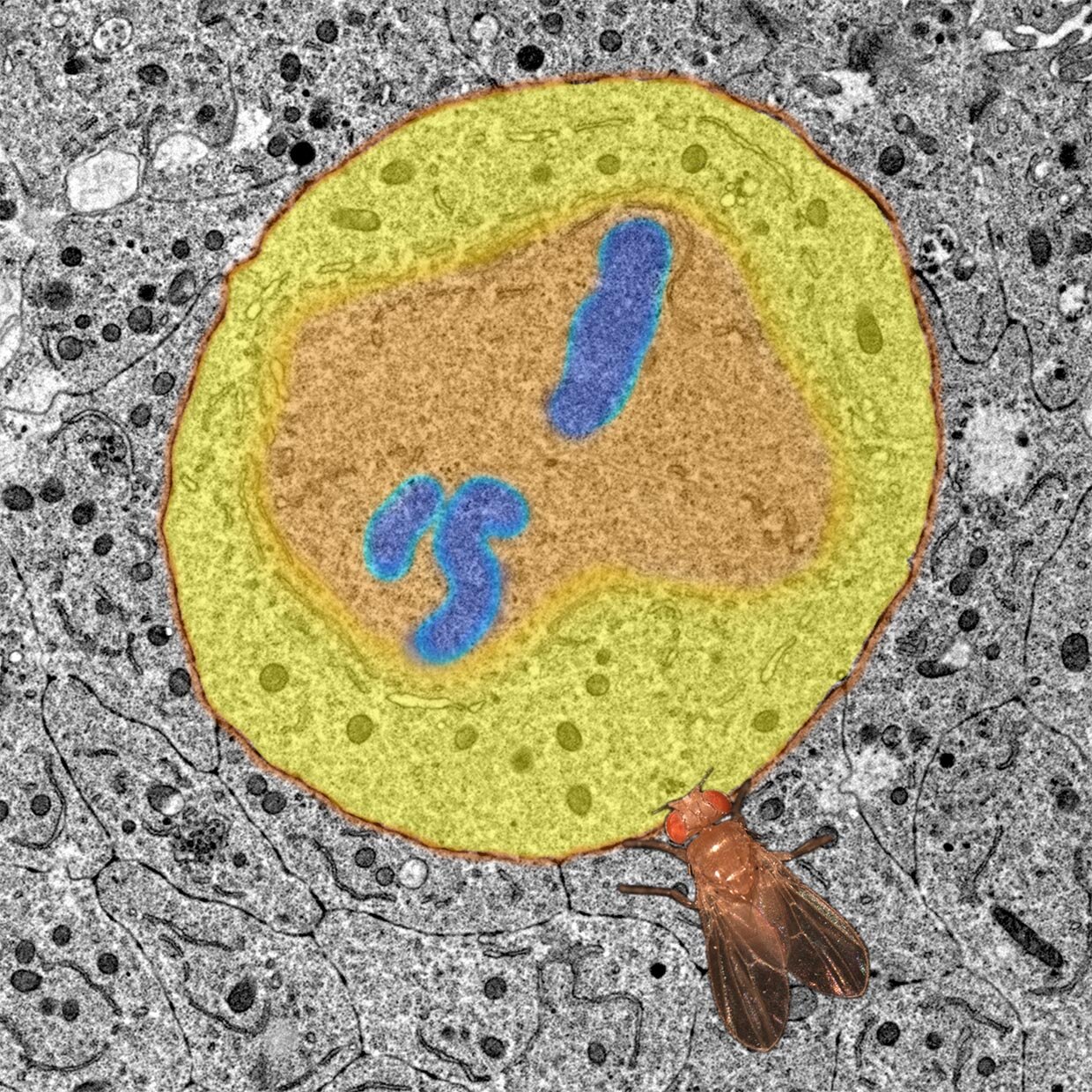
A Photoshop collage of transmission electron microscopy micrograph pseudo-coloured to highlight a dividing Drosophila melanogaster wing imaginal disc cell (x3000). A dividing cell of a developing wing tissue with a fruit fly observing the miracle of cell division.
FR: Un collage d’images de microscopie électronique à balayage colorisées pour mettre en évidence une cellule de l’aile de la mouche Drosophila melanogaster (magnification x3000). On peut ainsi observer le miracle de la division cellulaire.

Flower within a Flower
Robertas Ursache | Department of Plant Molecular Biology, UNIL, Switzerland
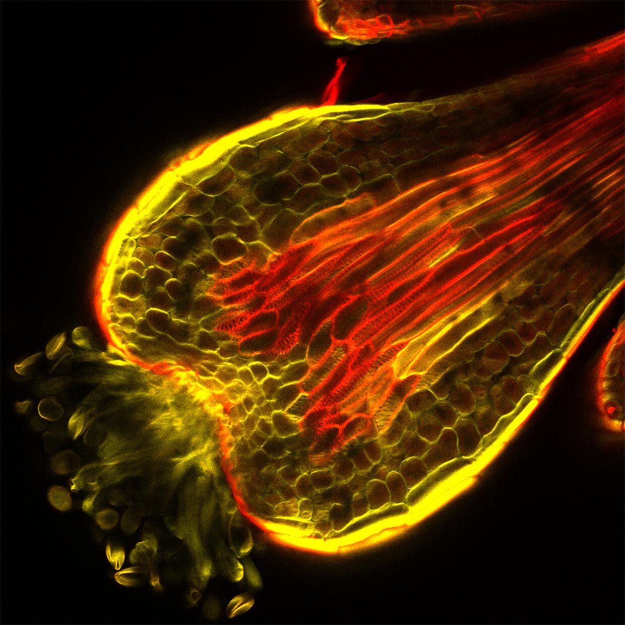
The stigma, a part of the flower of Arabidopsis thaliana where the pollen lands and starts the fertilization process. The image comes from a study on how the plant delivers water to these specialized structures. As revealed by Basic Fuchsin staining (in red), the water-conducting tissues form an interesting structure resembling a watering can.
FR: Le stigma, une partie de la fleur d' Arabidopsis thaliana où le pollen se situe et où commence le processus de fertilisation. L’image provient d’une étude sur comment la plante fournit l’eau à ces structures spécialisées. Comme l’a révélé par coloration de fuchsine basique (en rouge), les tissus conducteurs d’eau forment une intéressante structure ressemblant à un arrosoir.
AUDIENCE FAVOURITES

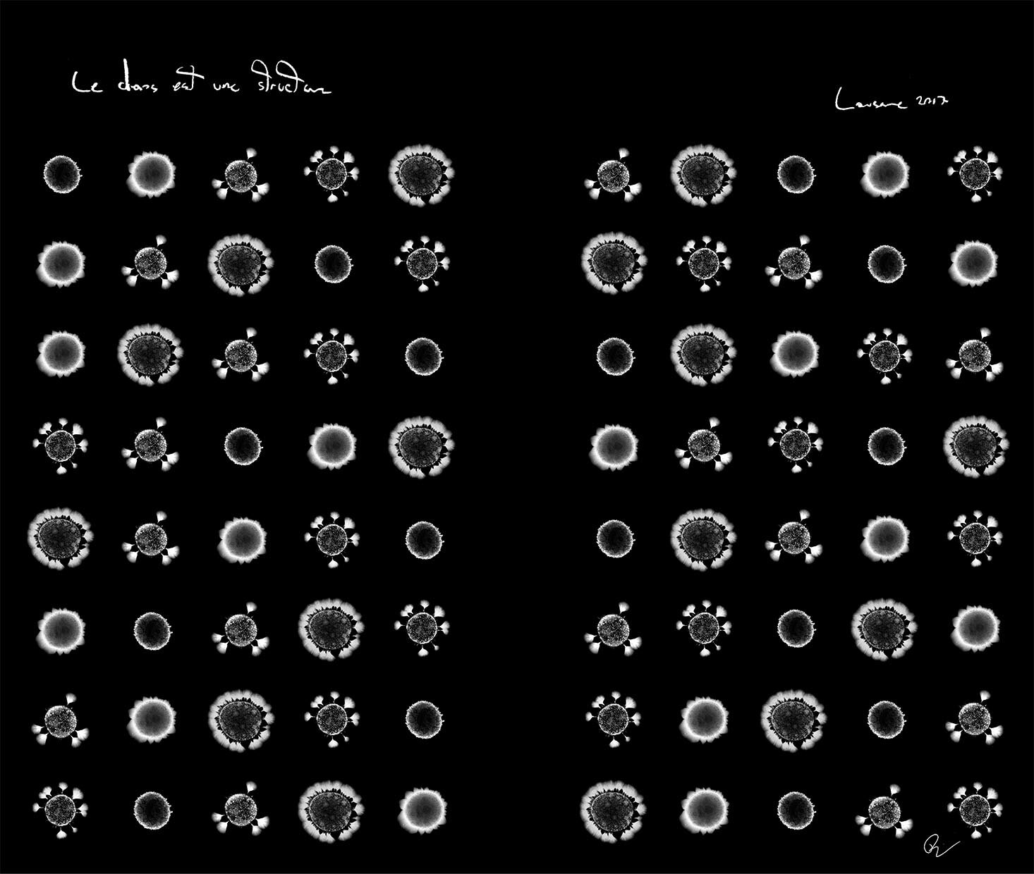
Le Chaos est une structure
Philippe Piccardi | Department of Fundamental Microbiology, UNIL, Switzerland
Microscopic images of colonies of the microorganism Agrobacterium tumefaciens (in white) mixed with other microorganisms growing on agar plates. The different colonies show that diverse growth patterns can emerge in a simple experiment. The random distribution but orderly arrangement of the five pictures (repeated sixteen times) is an allegory of the interplay between randomness and determinism in the organization of living systems. The human mind - with its limited perception - cannot make sense of this chaos, except with approximate predictions.
FR: Images microscopiques de colonies du micro-organisme Agrobacterium tumefaciens (en blanc) mélangées à d'autres micro-organismes poussant sur des plaques de gélose. Les différentes colonies montrent que divers modèles de croissance peuvent émerger d'une expérience simple. La distribution aléatoire mais la disposition ordonnée des cinq images (répétée seize fois) est une allégorie de l'interaction entre le hasard et le déterminisme dans l'organisation des systèmes vivants. L'esprit humain - avec sa perception limitée - ne peut donner un sens à ce chaos, si ce n'est par des prédictions approximatives.

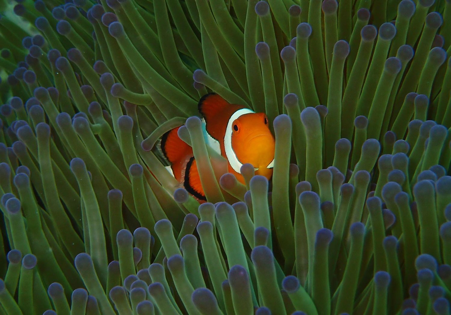
SYMBIOSIS
Sara Heim | Metabolimisc Unit, UNIL, Switzerland
The symbiotic mutualism observed between clownfish and sea anemone is believed to have triggered the adaptive radiation in clownfishes. Nevertheless, the reasons for which clownfish are able to remain unharmed by the stinging nematocysts are yet to be elucidated. The mucus coating present on clownfish skin may contain specific compounds allowing the interactions with the sea anemone and it might be the main element endowing immunity to these animals from their host. For this reason, the metabolomics analysis of clownfish mucus could provide precious information on how this symbiotic mutualism established.
FR: Le mutualisme symbiotique observé entre le poisson-clown et l'anémone de mer est censé avoir déclenché la radiation adaptative chez les poissons-clowns. Néanmoins, les raisons pour lesquelles les poissons-clowns sont capables de rester indemnes des nématocystes urticants restent à élucider. Le mucus présent sur la peau des poissons-clowns pourrait contenir des composés spécifiques permettant les interactions avec l'anémone de mer et pourrait être le principal élément conférant à ces animaux une immunité vis-à-vis de leur hôte. Pour cette raison, l'analyse métabolomique du mucus du poisson-clown pourrait fournir des informations précieuses sur la façon dont ce mutualisme symbiotique s'est établi.

This work is licensed under a Creative Commons Attribution-NonCommercial-NoDerivatives 4.0 International License.